fbpx

Five digital imaging systems...

…used in the Plant Phenotyping and Soil Health Facility


The Plant Phenotyping and Soil Health Facility at Cranfield University facility has the capability to replicate the entire cropping cycle, from tillage to post-harvest, both above and below ground, in a controlled glasshouse environment. Additionally, these facilities are integrated with Agri-EPI’s multi-sensor gantry-based phenotyping platform.

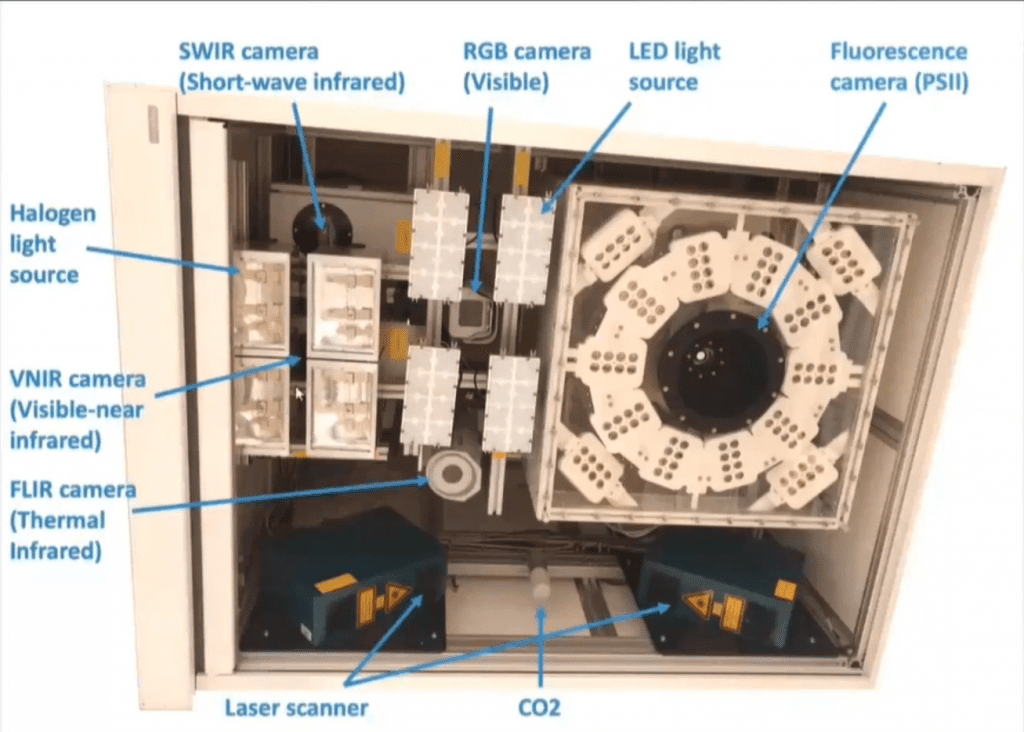
Bridging the gap between lab-based academic research and in-field research is a key challenge in crop health studies. To this end, the glasshouse at Cranfield benefits from an integrated automation system (with temperature and ventilation control), enabling a precise control for such variables in studies. The bespoke digital phenotyping gantry mounted inside the glasshouse is equipped with a large box containing a suite of six sensors. This box can easily be manoeuvered at precise locations throughout the glasshouse and an approximate 3.5m height adjustment can also be reached to accommodate analysis of small crops grown on benches. It can equally be readjusted for taller crops such as maize which the glasshouse can easily fit due to its 11-metre height. The sensors use a range of digital phenotyping technologies to monitor plant development.


1. RGB camera – visible light imaging

One of the sensors in the gantry is a high-resolution visible RGB camera, which is accompanied by a set of LED lights to provide optimal lightning. As a result, this technique produces very high-resolution images at the spectral bands of visible light (RGB, 400-750 nm). The benefits of this range from tracking  and assessing shoot biomass, plant traits, seed morphology, food quality and safety (among others) in controlled environments, to canopy cover in field studies

2. Lidar – sensor scanner

Sitting next to an equipped CO2 sensor is the Lidar (Light Detection and Ranging) scanner. In fact the gantry has two Lidar scanners, both fitted at 30 degree angles. These steeply angled near-infrared (NIR) laser scanners enable the generation of extremely detailed 3D images of different angles of plants (0.25 mm spaced points in all three axes). They are routinely used to study and characterize plant structure or soil surfaces.

3. FLIR IR Camera – thermal imaging

Placed right in the middle of the system is the thermal infrared camera. This imaging system is used to produce thermal images of plants, whereby changes in temperature of targeted plant components or areas can be detected. On a broader scale, thermal imaging can be used to investigate crop-water relationships, changes induced due to biotic or abiotic stresses and breeding programmes.

4. Hyperspectral cameras

To the left of the system, there are two types of hyperspectral cameras, with halogen lightning.

  • Visible-near infrared (VNIR) hyperspectral camera
    This operates in the visible and near-infrared range of the electromagnetic spectrum (380-1000 nm) and is used to detect spectral changes in the physiology and composition of crops produced by biotic or abiotic stresses.
  • Short-wave infrared (SWIR) hyperspectral camera
    Operating in the shortwave infrared range of the electromagnetic spectrum (950-2500 nm,the SWIR camera can also detect specific cellulose, lignin and leaf-water related indices.


5. Fluorescence imaging camera – PSII efficiency

The biggest sensor fitted in the system is the fluorescence sensor, which is surrounded by an array of LEDs that shine a high-intensity red light onto a plant. The small fluorescence measurement is carried out by the sensor in the system, which is directly linked to the photosynthetic activity in the plant. Fluorescence is light produced by a substance that has absorbed light or other radiation, which has a longer wavelength than the light that has been absorbed. Exposing the plants to continuous light induces a variation in the fluorescence of the crops, which allows chlorophyll fluorescence to be measured (based in Photosystem II. This system is often used as a tool to characterize and screen photosynthetic samples. It produces parameters that can be used of the early detection of diseases, such as bacteria on courgettes.

The imaging techniques discussed above allow us to push the limits of existing plant science to advance the detection of changes induced by external factors, such as pests and diseases or water availability, or internal factors, resulted from plant breeding. By using this imaging techniques in combination with Artificial Intelligence technology we can develop cost effective sensors that can be installed in mobile applications like drones.


Find out how CHAP can help to take your crop research to another level, go to Plant Phenotyping and Soil Health Facility and  Digital Phenotyping Lab.

Reach out to the CHAP team to discuss potential project opportunities using our Digital Phenotyping Lab: email us using the enquiries form at the bottom of our homepage.